
Currently all of the MCZ fish holdings can be searched directly through MCZbase, VertNet, iDigBio and GBIF.
Global Distribution of MCZ Fish Collection Localities


The Fish Imaging Project began in 2001 and since then over 2800 specimens have been photographed and posted on our searchable online database. The emphasis of this project has been photographing primary type specimens to facilitate examinations by researchers around the world. As of Feb 2013, an estimated 95% of Holotypes, Neotypes and Lectotypes in the collection have been photographed. Over the years, a number of department staff worked on the digital imaging project, including Chris Kenaley, Anne Holmes, and currently Andrew Williston and Karsten Hartel.
Major changes to the imaging project have taken place over the last six years. Equipment upgrades to our cameras, microscopes and computers have tried to keep pace with the explosion of digital imaging technology. Labels accompanying type specimens are now being scanned to preserve supplementary data.
Improvements in the MCZ Digital Imaging Facility have expanded the Fish Department's digital imaging capabilities. Our digital x-ray facility has allowed us to greatly expand our collection of radiographs. A Snycroscopy Auto-Montage system allows us to take images of our smallest specimens with almost unlimited depth of field. Now, the addition of a micro-CT scanner improves our ability to non-destructively image internal anatomy. This growing range of imaging equipment at the MCZ will greatly contribute to the growth of our digital media archives.
 The Camera
 The Wet Box
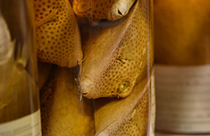
Many of the specimens we image are delicate and very susceptible to desiccation. To protect the specimens from the very intense heat of the lamps we developed a glass bottomed wet box (Figure 1C, 2). The wet box sits on the base of the copy stand and the specimen sits in fluid in the wet box.
In addition to its protective qualities, the light box allows the specimen to sit in a fluid media whereby delicate structures such as fin ray elements or escae can float freely. The detail and sharpness this method produces is often stunning. The walls of the wet box are made of inch plexi-glass and are joined by acrylic cement. The glass bottom sits in a sealed dado. The legs of the wet box lift the unit 3 inches from the base of the copy stand; elevating the stand leaves the background of the fish out of focus, providing an even backdrop.
 The Scope Mount
Small specimens between 0-15 mm usually require some magnification beyond a macro lens. In these cases we photograph using a Leica MZ9 dissecting scope fitted with an 8MP Nikon Coolpix 8400. Our camera is also connected via video outputs to a Trinitron monitor. This monitor allows multiple researchers to examine and discuss the specimen under the microscope.
The D5000 is directly interfaced with a dedicated desktop computer using Nikon Capture Control Pro 2 software. Capture control allows the user to adjust many of the relevant camera settings and take pictures with just the click of a mouse. Photographs are automatically downloaded through a USB connection. Editing to erase dust particles in the fluid or on the background is done in Adobe Photoshop CS5. Images are archived according to a museum-wide protocol, wherein original, untouched files are retained as raw and DNG files. Edited, publication quality images are saved and archived as .tiffs. Final images (as .jpeg) are linked to specimen records in the museum database.
The MCZ Larval Fish Archive is an ongoing project that curates and houses specimens of larval fishes for research use by the scientific community.
The archive had its beginnings in the transfer of the fish collections at the Woods Hole Oceanographic Institution to the Museum of Comparative Zoology in the late 1970s. It was found that a large part of these collections were valuable larvae and small juveniles of epipelagic and mesopelagic species. With support from the National Science Foundation (BSR85-01268 and 86-17845), S.L. Richardson and then D.G. Smith were brought to the MCZ to sort and identify these specimens and to establish a larval archive. This preliminary phase of the was completed by the late 1980s and new material is constantly being added during a second phase.

The Larval Fish Archive now has some 28,000 lots (141,000 specimens). The bulk of the archive is from the Woods Hole Oceanographic Institution. Additional material from the Gulf of Maine (H. DeWitt), the Bahamas (P. Major), the Coral and Solomon seas and world-wide blue water SCUBA collections (G.R. Harbison), and Mid-Atlantic Bight voucher specimens (M.P. Fahay) greatly supplement this Atlantic pelagic collection.
One of the goals of this project is to disseminate the information and to stimulate the use of this resource. All of this material is available for loan to recognized institutions.
We urge researchers to deposit voucher material from their larval studies in a permanent archive. The MCZ Larval Fish Archive has been designated a national repository and we have the responsibility to house voucher material from all areas of ichthyological research. We accept figured specimens, unique specimens, transforming juveniles, and developmental series either raised in aquaria or collected from the wild. We also accept voucher material from faunal surveys but may not be able to accept thousands of lots from any one locality.
Selected images are available through MCZbase.
|

 |
 |